Robotic Partial Nephrectomy - Single Port and Multi-Port
A kidney-preserving operation for most small and moderately sized kidney tumours - the tumour is removed with a thin rim of healthy tissue and the rest of the kidney is kept intact.
What is a partial nephrectomy?
A partial nephrectomy (also called a "nephron-sparing" operation) removes a kidney tumour together with a small margin of surrounding tissue, leaving the rest of the kidney in place and working. The operation is performed through small keyhole incisions using a surgical robot, which gives Dr Kooner magnified 3D vision and very fine, articulated instruments to work with.
For most renal masses under about 7 cm - and often for larger tumours depending on position - partial nephrectomy is the preferred surgical approach in current international guidelines.
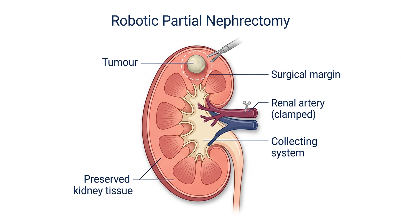
Why kidney-preserving surgery matters
- Equivalent cancer outcomes compared with removing the whole kidney, when the tumour is suitable for partial removal.
- Better long-term kidney function - preserving working kidney tissue lowers the lifetime risk of chronic kidney disease and dialysis.
- Cardiovascular benefit - chronic kidney disease is linked to heart disease, so keeping kidney function helps the whole body.
- Reserve for the future - if a new tumour develops years later, the remaining kidney tissue may itself need protecting.
Who is it for?
Partial nephrectomy is generally considered when:
- A solid kidney lesion is confirmed on imaging and is technically resectable.
- The patient has two working kidneys (though partial surgery is even more important when there is only one).
- There is no evidence of widespread disease on staging scans.
- The patient is fit enough to undergo robotic abdominal surgery.
Selection depends on tumour size, position within the kidney, proximity to blood vessels and the collecting system, and overall kidney function. Some complex tumours are best treated with a full nephrectomy; Dr Kooner will go through this honestly at consultation.
How Dr Kooner performs it
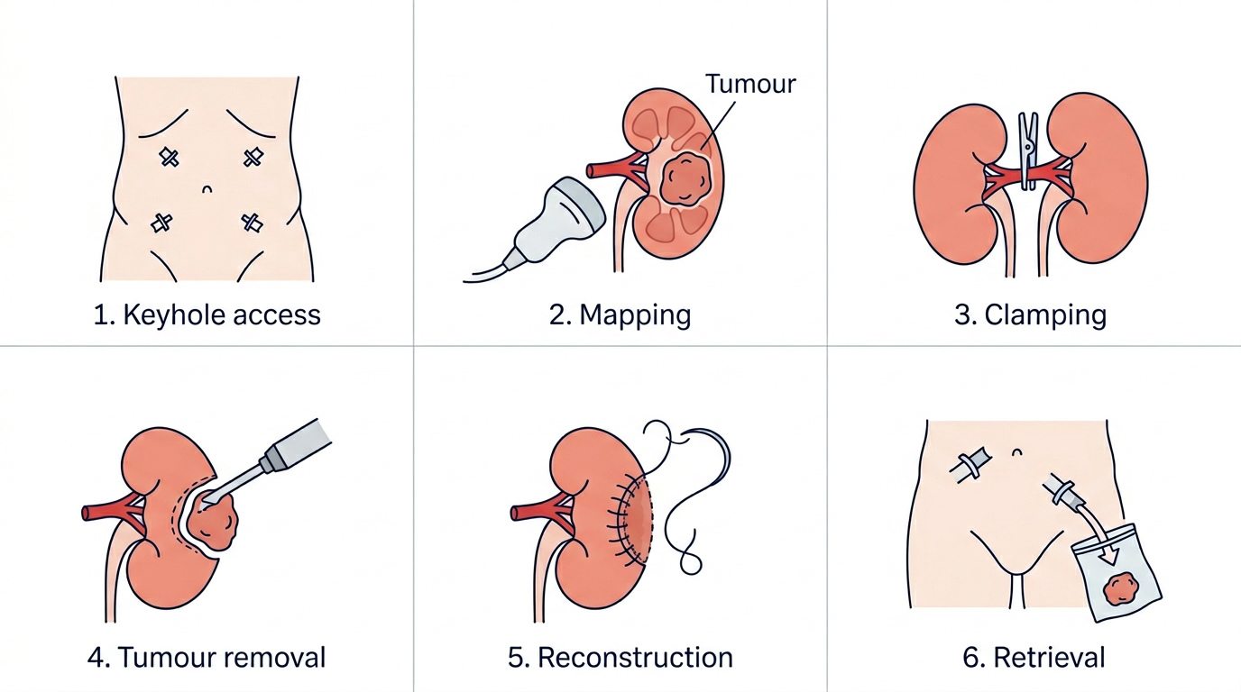
Robotic partial nephrectomy is a precise, technically demanding operation. The typical steps are:
- Access: four or five small keyhole incisions are made in the abdomen or flank (multi port robot). Alternatively, a single small incision is made (single port robot).
- Mapping: the kidney is carefully mobilised and the tumour identified under 3D vision, often with real-time ultrasound.
- Clamping (if needed): the blood supply to the kidney is briefly paused so the tumour can be excised in a bloodless field.
- Tumour removal: the tumour is cut out with a narrow margin of normal tissue.
- Reconstruction: any opened collecting system is closed, and the kidney is reconstructed in layers with fine sutures to restore its shape and integrity.
- Retrieval: the tumour is placed in a specimen bag and removed through one of the small incisions.
Goals of the procedure
Dr Kooner operates to the "trifecta" standard routinely described in partial nephrectomy literature: complete tumour removal, preserved kidney function, and no significant complications.
- Negative margins on final pathology (no tumour left behind).
- Minimal warm-ischaemia time while the blood supply is paused.
- Preserved long-term kidney function, measured on follow-up blood tests.
Your journey - what to expect
-
Consultation & planning
Review of CT/MRI imaging, baseline kidney function tests, anaesthetic assessment, and a clear discussion of options, risks and expected recovery.
-
Admission & theatre
Admission on the morning of surgery. The operation is performed under general anaesthetic and typically takes two to four hours, depending on tumour complexity.
-
Hospital stay
Most patients are up walking the day after surgery, eating normally within 24–48 hours, and home within 2–4 days with simple pain relief.
-
Recovery at home
Light activity from day one, driving and office work typically around 2 weeks, with a gradual return to exercise and heavy lifting by 4–6 weeks.
-
Pathology review & surveillance
Results are discussed 2–3 weeks after surgery. A tailored surveillance schedule (imaging and blood tests) is arranged based on the final histology and stage.
Risks & considerations
Partial nephrectomy is a major operation and, as with any surgery, it carries risks. Dr Kooner will go through these individually at consultation. The main ones to be aware of are:
- Bleeding, occasionally requiring transfusion or (rarely) a return to theatre.
- Urine leak from the reconstructed kidney, usually managed with a temporary stent.
- Infection, chest or wound complications - uncommon with keyhole surgery.
- A small reduction in kidney function on the treated side, usually offset by the opposite kidney.
- Conversion to open surgery or to removal of the whole kidney - uncommon but discussed as a possibility in advance.
- Risks related to general anaesthetic, DVT/PE, and the abdominal surgical field.
Frequently asked questions
Is partial nephrectomy as effective as removing the whole kidney for cancer?
Yes, for tumours that are suitable for partial removal. Long-term cancer outcomes are equivalent, and preserving kidney tissue gives better long-term health overall. Dr Kooner will explain whether your specific tumour is suitable.
Why is the robot used rather than ordinary keyhole surgery?
The robot allows very fine suturing deep in the abdomen, which is essential when the kidney has to be reconstructed quickly under a time pressure (the blood supply is paused). 3D magnified vision and wristed instruments make complex partial nephrectomies safer and more reproducible than standard laparoscopy.
Will I need dialysis afterwards?
For most patients with two normal kidneys, no. Partial nephrectomy is specifically designed to preserve function. Patients who start with reduced kidney function, or who have only one kidney, are assessed carefully and often benefit most from this kidney-preserving approach.
How long will I need off work?
Most people with desk-based jobs return to work around 2 weeks after surgery. Those in physical jobs usually need 4–6 weeks. These timings are a guide - recovery is individual.
What follow-up will I need?
Follow-up depends on the final pathology. For most kidney cancers this involves periodic imaging (CT or ultrasound) and blood tests for several years, stepping down in frequency over time.